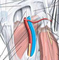
The PMTL is made up of the popliteus muscle, the popliteofibular ligament (PFL), the femoral insertion of the popliteus tendon, the popliteomeniscal fascicles and soft tissue attachments to the lateral meniscus, and the proximal tibia.
Click here to see the full labelled illustration.
The popliteus tendon and PFL are the most important elements of this unit in terms of providing stability to the knee. These structures aid the FCL and posterolateral capsule to prevent excessive external tibial rotation and varus rotation. [That is, they keep the bones stable in that corner of the knee.]
When the PMTL is deficient, it must be reconstructed with a graft (in addition to a graft replacment of the FCL) to fully restore posterolateral corner function.
The popliteus muscle originates at the back of the tibia and proceeds around the side and up (lateral and proximal) to insert on the lateral femoral condyle. Its tendon proceeds on proximally through a gap in the coronary ligament of the lateral meniscus, then passes deep to the FCL to ultimately insert in front of and below (anterior and distal) to the insertion of the FCL.
The PFL originates at the musculotendinous junction of the popliteus and attaches to the medial aspect of the fibular head, where it lies deep to the fabellofibular ligament.
[This article was published in Noyes’ Knee Disorders: Surgery, Rehabilitation, Clinical Outcomes, Noyes FR, Barber-Westin SD (eds.), Copyright Saunders, 2009 - Strickland JP, Fester EW, Noyes FR: Lateral, posterior, and cruciate knee anatomy, pages 20-43.]


