The two basic x-ray views are called the AP and Lateral. These are the front (anterior-posterior) and side views. These are the views obtained in an Emergency Room and they are useful for detecting major fractures, large tumors, and advanced arthritis.
Evaluation of the patella, though, usually requires special views.
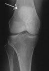
Take a look at this x-ray. The bones are pale, and the spaces are dark. If one bone overlies another you may see the outline of both, but they appear to be on the same plane (see arrow, showing the pale circle of the patella overlying the femur). With a single film one can thus see only the one dimension. This view is called an 'AP' view (antero-posterior).
In an AP view the patella shows as a vague circle over the femur bone. Little information can be gleaned other than its height and basic outline, and maybe fracture (break) lines (which appear dark).
An improvement on the basic AP view is the Standing AP view. Here you are standing as the x-ray is taken. If you suffer from knee arthritis, the narrowing of the space between the bones that is a sign of arthritis is best seen when you stand.
Another variation on the AP x-ray is the Standing Tunnel AP. This view also goes by the name Standing Flexion view, Rosenberg view, or even Schuss view because the patient stands with the knees bent. This view will occasionally detect arthritis that even the regular Standing AP will not pick up.
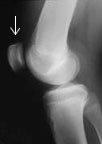
One needs generally to take another picture from the side to see the other dimension. This is called a 'lateral' view. Now you can see that femur and patella are in different planes.
The Lateral (side) view can be technically difficult. In order to get an x-ray that is in the correct plane, the x-ray technician may have to rotate the leg this way or that way until he or she gets it right. By always seeing a standard view doctors can develop confidence in interpreting the films.


