Successful arthroscopic surgery dictates that the surgeon view the joint from more than one angle.
First published by knee surgeon Angus Strover in 2008, and reviewed August 2023 by Dr Sheila Strover (Clinical Editor)
The value of the suprapatellar portal
Successful arthroscopic surgery dictates that the surgeon view the joint from more than one angle.<
First published by knee surgeon Angus Strover in 2008, and reviewed August 2023 by Dr Sheila Strover (Clinical Editor)
I have published this to my colleagues in a leading surgical journals, but still today I have professional surgeon visitors who are amazed when I demonstrate this to them.
I am talking about something quite simple, but surprisingly seldom performed - and that is simply to examine the joint during arthroscopy FROM ABOVE.
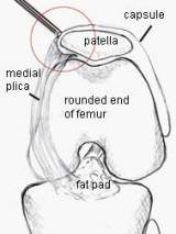
You will remember that so far I have taken you through the routine of starting with the arthroscope in the anterolateral portal (AL) and examining the medial side (as in the photo), then swapping it over to the anteromedial portal (AM) and examining the lateral side (link). From these two portals, much of the knee cavity can be viewed and probed.
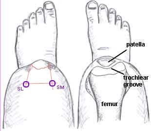
But switching portals yet again, this time to those ABOVE the patellar give the surgeon further very important information. These portals are called the suprapatellar portals - that is the superolateral (SL)) and the superomedial (SM) portals respectively. With the scope in these portals, the tip of the scope is in the cavity (suprapatellar space) above the patella, and one looks DOWN in the direction of the feet.
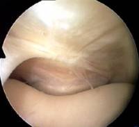

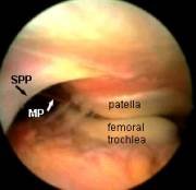
I've mentioned plicae already on this course. Although a plica is a normal structure, some people have them and some don't. They may differ in situation, size and thickness. If a medial plica is abnormal and thickened it can often be felt as a string-like object to the inner side (medial) of the patella. From the suprapatellar portal it is amazing how easy it is to see why medial plicae cause pain.

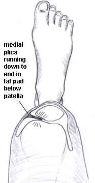
Look again at this region but this time from the front and side (all the left knee).
It should be clear that from the usual anterolateral portal one cannot see much of what is going on in this suprapatellar area because the bulge of the rounded femur gets in the way.
Certainly one cannot appreciate the plica being nipped between patella and femur in the way that you can from above.
The medial plica sweeps along the side of the patella, but the suprapatella plica lies horizontally above the patella,and may stretch right across the joint cavity (the 'suprapatellar pouch').
Here is an MRI scan, where the contrast has been reversed so that the bones are dark and the knee cavity light.
The yellow arrow is pointing to a suprapatellar plica, which you can see is stretched across the suprapatellar pouch above the patella.
This is why one must measure two fingersbreadth above the patella before inserting the arthroscope, or the scope may be too low and this plica may be missed.
Here the arthroscope has been withdrawn above the level of the medial plica to reveal the suprapatellar plica (black arrow). You can see its position in relation to the medial plica (white arrow), which is just draping itself out of sight over the front of the femur. REMEMBER, WE ARE LOOKING FROM ABOVE, WITH THE SCOPE IS THE LARGE CAVITY ABOVE THE PATELLA. You can't see this structure from the ordinary portals!
A suprapatellar plica may stretch right across the suprapatellar pouch, dividing it into two. This is called a 'complete septum', and its presence may confuse a novice arthroscopist. It is easier for you to understand a complete septum if you see an almost complete one - what we call a 'fenestrated' plica. That means 'a plica with a window'.
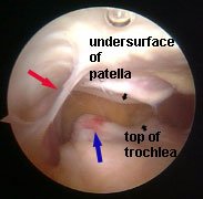
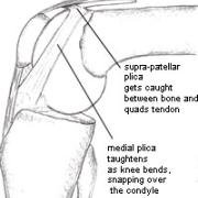
A plica may also be like a band of tissue. This image is of a suprapatellar plica viewed from the lateral suprapatellar portal, bowstringing and snapping across the supra patellar pouch over the medial femoral condyle. In this image, the red arrow is pointing to the plica. The little black arrows show the joint surface of the patella and femur.
The blue arrow shows the inflamed area of the femur where the plica has been snapping. This would be completely invisible from the lower portals.
Besides the advantages already mentioned, the supra-patellar portal allows -
I'm going to end this lesson at this point, but I just want to show you the video of patellar tracking. Click the arrow to start the video.