Careful evaluation is essential to determine how a posterolateral corner injury has occurred and which anatomical structures are damaged.
First published 2009 by Dr Frank R Noyes, and reviewed August 2023 by Dr Sheila Strover (Clinical Editor)
Mechanism of injury of the posterolateral corner
Careful evaluation is essential to determine how a posterolateral corner injury has occurred and which anatomical structures are damaged.
First published 2009 by Dr Frank R Noyes, and reviewed August 2023 by Dr Sheila Strover (Clinical Editor)
A blow (from an opponent or object) to the top and inside (anteromedial) part of the tibia during sports and motor vehicle accidents are common ways that the PLC is injured.
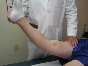
As well, a noncontact injury in which the knee hyperextends and the tibia twists outward may tear these tissues. High-energy trauma, such as that sustained in a motor vehicle accident, is another common cause of PLC injury.
An isolated complete PLC rupture is rare as usually the injury is accompanied by a tear to the ACL or PCL.
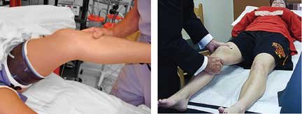
A comprehensive evaluation of the PLC, as well as all of the other major knee structures, must be performed to determine all of the injuries or deficient ligaments and soft tissues, as well as any abnormal alignment or bone problems. To exclude any of the tests described below may result in an improper diagnosis.
At my Center, we do the following:
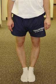
ie. knee joint bowing outwards and hyperextendable, that is bending backwards
ie. how far the kneecap moves side-to-side, crepitus on movement, pain
[This article was published in Noyes’ Knee Disorders: Surgery, Rehabilitation, Clinical Outcomes, Noyes FR, Barber-Westin SD (eds.), Copyright Saunders, 2009 - Noyes FR, Barber-Westin SD: Primary, double, and triple varus knee syndromes: Diagnosis, osteotomy techniques, and clinical outcomes, pages 821-895.]
MRI, bone scan: if required, done for multiple ligament injuries
X-rays
For dislocated knees, a lower extremity venous ultrasound is obtained in knees that have swelling and soft tissue damage. An initial delay before any surgery is considered for 5 to 7 days to allow for observation of the neurovascular status, soft tissue swelling, skin integrity, and some clearing of hemorrhage in soft tissues in the injured extremity.
It is important to understand that injuries to the PLC structures vary, with some patients sustaining only mild to moderate damage and others having severe ligament and soft tissue ruptures. The examination I just described allows the classification of these injuries into either a first, second, or third degree injury.
The treatment of these injuries depends on the degree of damage. First and second degree injuries do not require surgery, but are treatment with rehabilitation and bracing in some cases, as discussed in Part 4. The third degree injury patterns require much more extensive treatment, as I will discuss in Parts 4 and 5.


