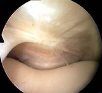
This arthroscopic photograph is looking down on the plica from above, and you can just see the top of the patella at the bottom of the image as it engages the groove of the femur.
Commonly a suprapatellar plica is a curved band along one wall of the capsule above the kneecap.
Rarely a suprapatellar plica may reach right across as a full membrane from one side of the supra patellar pouch to the other, dividing it in two, sometimes with a little window in the middle. Then it is called a suprapatellar septum. Although a septum can also be symptomatic, its real importance is that it often confuses the novice surgeon during arthroscopy.
Peer-reviewed papers
Quote from peer-reviewed paper:
"....The knee joint is believed to have formed in the eighth week of fetal life with three compartments....Then, the synovial septa are partially resorbed over the next several weeks, and a single joint cavity is created...[and] remnants of the unabsorbed membrane are recognized as synovial plicae"
Citation: Akao M, Ikemoto T, Takata T, Kitamoto K, Deie M. Suprapatellar plica classification and suprapatellar plica syndrome. Asia Pac J Sports Med Arthrosc Rehabil Technol. 2019 Apr 22;17:10-15. doi: 10.1016/j.asmart.2019.03.001. PMID: 31044135; PMCID: PMC6477514.
End of paper Quick links


