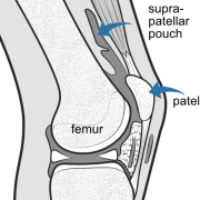
This illustration shows the knee cut through the middle. The large cavity (dark grey) extends above (suprapatellar pouch), behind and below the patella. A small suprapatellar plica is present but it does not form a complete septum so the whole cavity is open to the joint..
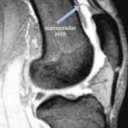
This MRI shows a similar view where the arrow is pointing to a complete suprapatellar septum dividing the cavity into two - the closed bursa (above) and a small pouch below. This may confuse the novice surgeon during arthroscopy.