
The position of the patella relative to the groove of the femur changes as the knee is bent. In the straight knee the patella lies above the groove and can be freely wiggled from side to side. As the knee is bent the patella engages with the groove of the femur and it should not be possible to wiggle it from side to side.
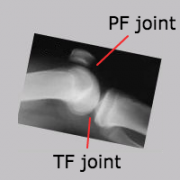
When you look at the X-ray again, you will see that the patella bone seems to be 'floating' - but it is just that the tendons above and below it do not show on X-ray. Neither do the two shock-absorber menisci, which appear just as a dark gap on X-ray between the two bigger bones.
Looking from above, when the knee is bent, the undersurface of the patella (kneecap) lies snugly like this in the trochlear groove. The patella should be central in the groove, and not favouring one or other side, and it should not be tilted. The sides of the patella and the walls of the groove should be almost parallel. The apparent gap that one sees on X-rays from this angle (Merchant views) is not really a gap but the space is filled with the white joint cartilage that covers both of the bony surfaces where they are in contact.
Note that you cannot see the rounded condyles of the femur in this view, nor can you see the tibia, as this angle looks down on the 'lap' and the condyles they are on the under-surface of the femur, while the tibia is at right angles to this view.
This view is also one that the surgeon can explore during arthroscopy, where he can get a superb opportunity to see how the patella glides in the trochlear groove. Many surgeons, unfortunately, miss this opportunity because they do not put the camera into this 'suprapatellar' region, and thus they fail to fully appreciate the anatomy here. This is also the best view to appreciate something called a 'plica', and problems with a plica may also be missed.
Back to top


