
So this group of prominent surgeons, including several leading Japanese surgeons, looked more carefully at the
anatomy and doing a different kind of
ACL reconstruction and came up with this concept of ‘double-bundle’
ACL reconstruction, as opposed to a single bundle
ACL reconstruction. That is a very anatomical way of doing the surgery as it is re-creating the
anatomy by replacing the two ruptured bundles of the original
ligament with two new bundles. The problem with that is that it is technically very difficult, and drilling two tunnels in the
tibia and two tunnels in the
femur is not only technically difficult but revising those patients if there is a problem can be a real challenge. there are so many problems with it that it has been pretty much abandoned and is no longer considered to be a mainstream way of doing
ACL surgery.
Last April (2009) a UK surgeon, Tim Spalding, launched his ideas at BASK with an excellent presentation last April (2009) in Oxford where he demonstrated an improved method of locating the real anatomical position for a single bundle procedure – what they are calling the direct measurement technique for ACL surgery. He has now followed that up with a clinical study with high-resolution CT scanning and that article has just been submitted for publication. The new ‘buzz’ way of describing an anatomical ACL reconstruction is called ‘footprint’ ACL surgery or ‘anatomical’ ACL surgery. Tim Spalding and his group have now done a very in-depth study looking at this work over the last year and they have found a very high degree of accuracy between the direct measuring technique that they developed correlated with CT scans to show that they have had a great success in putting the ACL exactly where it should be.
I have been working on a new technique that facilitates the placing of the new ligament in this real anatomical position – doing all the work from the medial side, which is much easier than the old techniques. So you actually look from the medial side and do all the work from the lateral side with some special instruments that go around the corner into the notch and allow you to prepare the femur, mark the femur and then accept this drill bit.
I would like to show you this method which I have illustrated via live surgery in Garmisch, in Southern Germany in 2011 (location of 2011 World Ski Championships) when I was presenting my approach to the German arthroscopic association, and you will find the video below.
This is the view from the patient’s room!

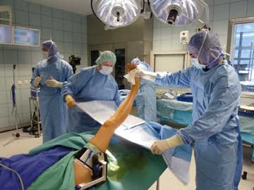
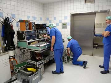
Here are two photos - one showing the surgical staff preparing the patient for surgery while the other shows the recording team seeing to the video and sound equipment.
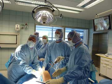
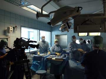
The next two photos show the surgical team in action looking up at the monitor with the arthroscope on the medial side - you can see how busy it all is with the video crew and the surgical team all in the room together!
Finally, I will show you a video of the new procedure if you click this link - http://www.youtube.com/watch?v=vKVcibPApPU.
