Dr Angus Strover explains the steps a knee surgeon will take in performing an arthroscopy.
First published by knee surgeon Angus Strover in 2008, and reviewed August 2023 by Dr Sheila Strover (Clinical Editor)
Diagnostic Arthroscopy - The Importance of Method
Dr Angus Strover explains the steps a knee surgeon will take in performing an arthroscopy.
First published by knee surgeon Angus Strover in 2008, and reviewed August 2023 by Dr Sheila Strover (Clinical Editor)
This is an important subject.
Why? Well, during my years as a knee surgeon, I had a great many patients come to see me because they were not improved after previous arthroscopic surgery. I was surprised in many cases to find that their previous surgeon had failed to locate the obvious source of their symptoms, and I realised that this was because the surgeon had simply not performed a thorough and systematic examination during the arthroscopy.
For over twenty years now and together with a number of dedicated colleagues, I have been trying to spread the use of a systematic method to the hundreds of young surgeons who attend our skills workshops at The Knee Foundation.
This course is aimed at an 'intermediate' level, and may be of interest to both patients, junior doctors and operating theatre staff.
This is what we are going to cover during the course:
Note that, except for a brief overview in the final lesson, we are not going to talk about the surgical procedures which a surgeon might perform during an arthroscopy - this is the stuff of more complex courses. The purpose of this current course is to show how important method is in arthroscopy - and how one can miss the diagnosis by using faulty method.
Something I want to impress on you is that the surgical routine does not start when the surgeon puts the knife to the patient's skin. It starts when the surgeon and patient agree that surgery will be undertaken. Time taken here to explain the procedure to the patient will save much of the anxiety patients may suffer in the build up to the actual procedure.
In my practice, I have a really useful three-dimensional model of the knee, and I take every patient through their procedure using this model. It is made by Adam Rouilly and I have found it over the years to be virtually indestructible. This little model is so useful that I give one to each of my departing knee fellows (trainees) when they head off back to their own countries to set up practices there!
The model allows a discussion of various knee cap problems such as tilt and maltracking; it helps to demonstrate the effect of procedures to improve the mechanics of the knee cap; the common sites of arthritis can be identified and relevant procedures for arthritis can be discussed; the cruciate ligaments are easily identified, and the procedures for reconstruction can be explained. And so on.
I think that this model is much more useful than pictures, as the two-dimensional nature of an illustration may cause the patient some confusion - whereas the model allows for a much more sophisticated level of dialogue altogether.
The term 'informed consent' should not mean that the surgeon has informed the patient what surgery he is going to undertake. It should mean that the patient should be an informed patient. The patient must understand -
The period just before the patient receives the anaesthetic is an important time, and again here a surgeon can miss an important opportunity to -
Picking up that last point, how easy do you think it is to do an arthroscopy on the wrong knee?
Yes. Terrifyingly easy.
Firstly, because the two knees might both look perfectly normal at the time, as there may be no swelling or external sign of anything wrong with the knee.
The patient is asleep when the surgeon is scrubbing. Also the nurse is draping the knee while the surgeon scrubs, and the surgeon generally accepts that the nurse won't made a mistake!
But a big black arrow on the bad knee, put there by the surgeon himself before the patient is put to sleep, ensures that such a disaster does not happen.
I won't go into detail about the anaesthetic, or discuss whether one should have this or that anaesthetic, but I would just like to tell you about one or two steps which I believe make a great difference to the patient.
I had the good fortune to work closely with a team of anaesthetists (anaesthesiologists) who were expert at doing what is known as 'regional blocks'.
A regional block is a nerve block with local anaesthetic, which blocks the nerve in its lower distribution, in contrast with a local block, which paralyses the nerve structures just around the injection itself. Some anaesthetists (anaesthesiologists) are good at this and are able to produce complete loss of sensation in the knee.
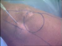
For ordinary arthroscopic procedures we used this for some time, but found that many patients were frustrated that they could not use their quadriceps muscles for sometimes several days afterwards.We now use local anaesthetic in the skin at the sites of the portals before the operation starts and also leave some local anaesthetic in the joint at the end of the procedure.
Here you can see the bulge in the skin where the local anaesthetic is being injected.
Don't worry about those pen marks. You will understand what they are when you get to a later section of the course.
A tourniquet is an inflateable wrap, which goes around the top of the thigh and gets pumped up to stop the blood flowing to the limb.
Probably the majority of knee surgeons apply and inflate a tourniquet before commencing the arthroscopy, and in addition the scrub nurse usually first exsanguinates the leg (squeezes the blood out using an elastic bandage). This offers the surgeon an advantage in there being virtually no bleeding during the procedure, but I believe this to be a two-edged sword for the following reasons:
For a routine arthroscopy I do not even apply a tourniquet!
Instead I make a point of controlling bleeding during the arthroscopy by these methods:
For beginners in this technique of arthroscopy it may be practical to apply a tourniquet but not inflate it. It can then be used if necessary. I started this way and graduated to the position of no tourniquet after a period of three or four years.
OK. This is the end of Part 1. In Part 2 I will discuss the basic instrumentation and its limitations.